KNEE | RENTON & MAPLE VALLEY
Patellar Instability and MPFL Reconstruction
Patellar instability is a condition in which the kneecap slips out of its groove, often tearing the MPFL ligament. Recurrent dislocations are commonly treated with MPFL reconstruction, sometimes combined with procedures that correct underlying alignment.
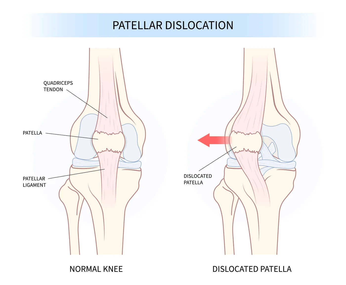
A kneecap dislocation is violent and memorable: the knee buckles, the patella visibly shifts, and the joint swells impressively. For some, it is a one-time event. But for many, especially teenagers and young adults, the first dislocation is the beginning of a pattern, because the initial injury tears the medial patellofemoral ligament (MPFL), the checkrein that holds the kneecap centered. Each subsequent dislocation risks shearing off cartilage, which is why recurrent instability is worth treating rather than enduring.
Why Kneecaps Dislocate
Some knees are simply built with more dislocation risk: a shallow groove (trochlear dysplasia), a kneecap that sits high (patella alta), or alignment that pulls the patella outward. Add a sports pivot or a direct blow, and the MPFL tears as the kneecap escapes the groove. Understanding each patient’s specific anatomy, through exam, X-rays, and often MRI or CT with standardized measurements, is the foundation of good treatment, because the operation should match the anatomy.
Treatment After a First Dislocation
Most first-time dislocations without a loose cartilage fragment are treated nonoperatively: a short period of protection, then dedicated therapy to rebuild quadriceps and hip control. Surgery after a first dislocation is generally reserved for cases with a loose body, a cartilage injury needing treatment, or anatomy that makes recurrence very likely. This is also the visit where risk gets discussed honestly, because recurrence rates in young patients with risky anatomy are substantial.
MPFL Reconstruction
For recurrent instability, MPFL reconstruction rebuilds the torn checkrein using a small tendon graft anchored between the kneecap and femur. It restores the passive restraint that keeps the patella tracking in its groove. When imaging shows significant underlying malalignment, Dr. Lyons may recommend combining the MPFL with a tibial tubercle osteotomy, repositioning the attachment of the patellar tendon, or addressing the groove itself, so that the reconstruction is not asked to fight the skeleton for the rest of your life.
Recovery
MPFL reconstruction typically allows early motion and weight-bearing in a brace, with progressive strengthening over the following months and return to cutting sports once strength and control criteria are met. Most patients describe the biggest change simply: the knee stops feeling like it is about to go.
Frequently Asked Questions
What is the MPFL?
The medial patellofemoral ligament is a band of tissue connecting the inner kneecap to the femur. It is the primary restraint against the kneecap slipping outward and is torn in most first-time dislocations.
Does a dislocated kneecap need surgery?
Usually not after a first dislocation, unless a fragment of cartilage or bone is loose in the joint. Surgery is typically recommended for repeat dislocations or high-risk anatomy.
What is MPFL reconstruction?
MPFL reconstruction replaces the torn ligament with a small tendon graft, restoring the checkrein that keeps the kneecap centered in its groove. It is the workhorse procedure for recurrent patellar instability.
Why would I need a bone procedure too?
If your anatomy pulls the kneecap outward, a soft tissue reconstruction alone may be overworked. Repositioning the tibial tubercle corrects the underlying pull so the new ligament can succeed.
If your kneecap has dislocated more than once, the pattern is unlikely to break on its own. Request an evaluation in Renton or Maple Valley.
Take the Next Step
Request an appointment at the Renton or Maple Valley office.